
Wisdom Teeth Development
Wisdom teeth are uniquely troublesome because they are the most frequent teeth to be partially or fully impacted (fail to erupt completely) with only 16% erupting normally. 1 This makes them extremely prone to trapping food and holding bacteria around their crown and root surfaces beneath the gum line. As a result, wisdom teeth are – by far – the single most likely teeth to have pathology associated with them and require repeated, expensive interventional treatment later in life at a rate of nearly 99%. 2, 3, 4, 5, 6, 7, 8, 9, 10, 11, 12 . The panographs below depict many of the problems commonly associated with wisdom teeth in adults.
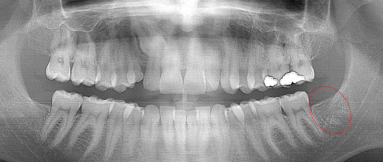
This 29 year old patient had only 1 third molar. It was extracted several years ago (extraction site circled). The outline of the roots remain evident. This patient had good post-op healing and an excellent boney contour develop. Unfortunately, this level of bone reformation does not always occur.
This 34 year old patient has all four third molars present (circled) and fully erupted into occlusion. They appear disease free…but are difficult to keep clean. 3rd molars are the most likely teeth to decay or have gum disease with a >98% probability that decay and gum disease will occur around all four teeth over this patient’s life time.
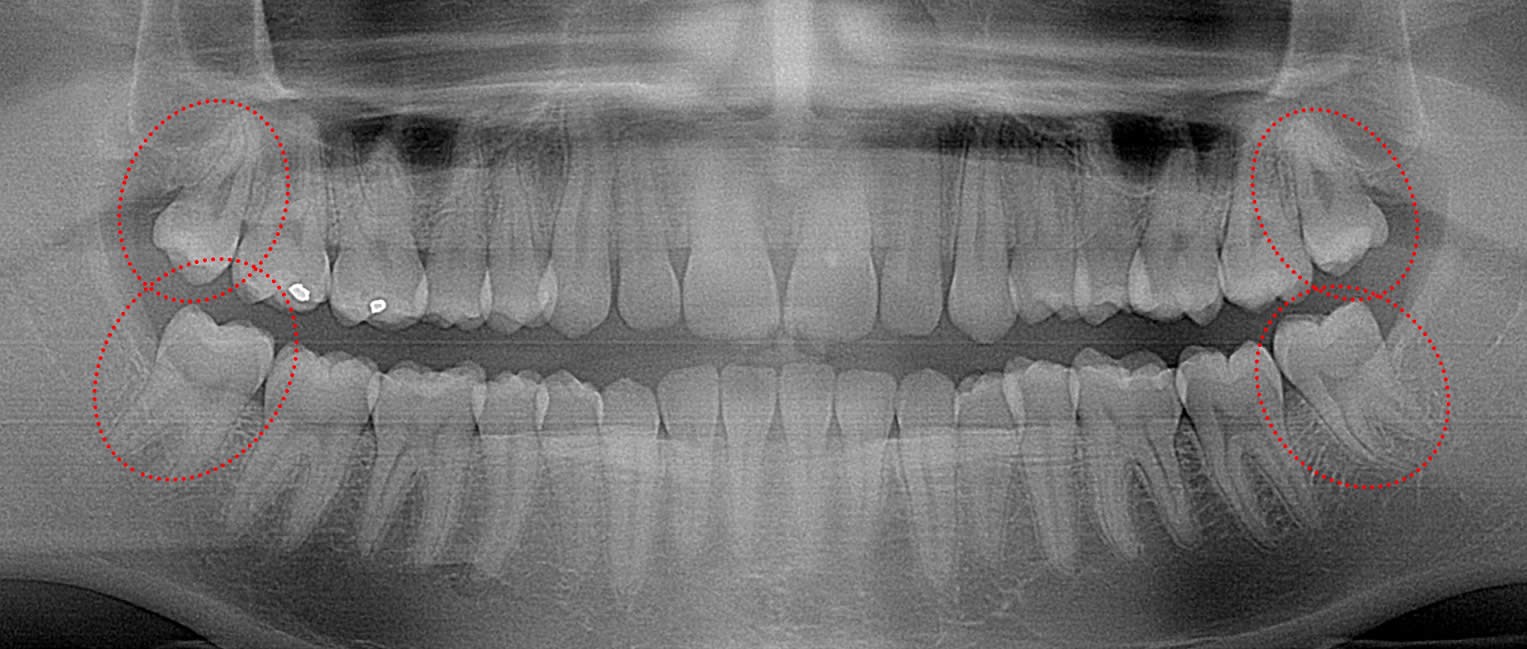
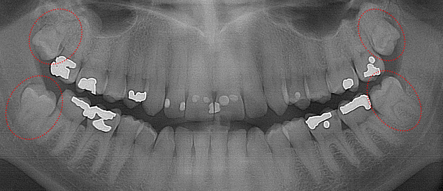
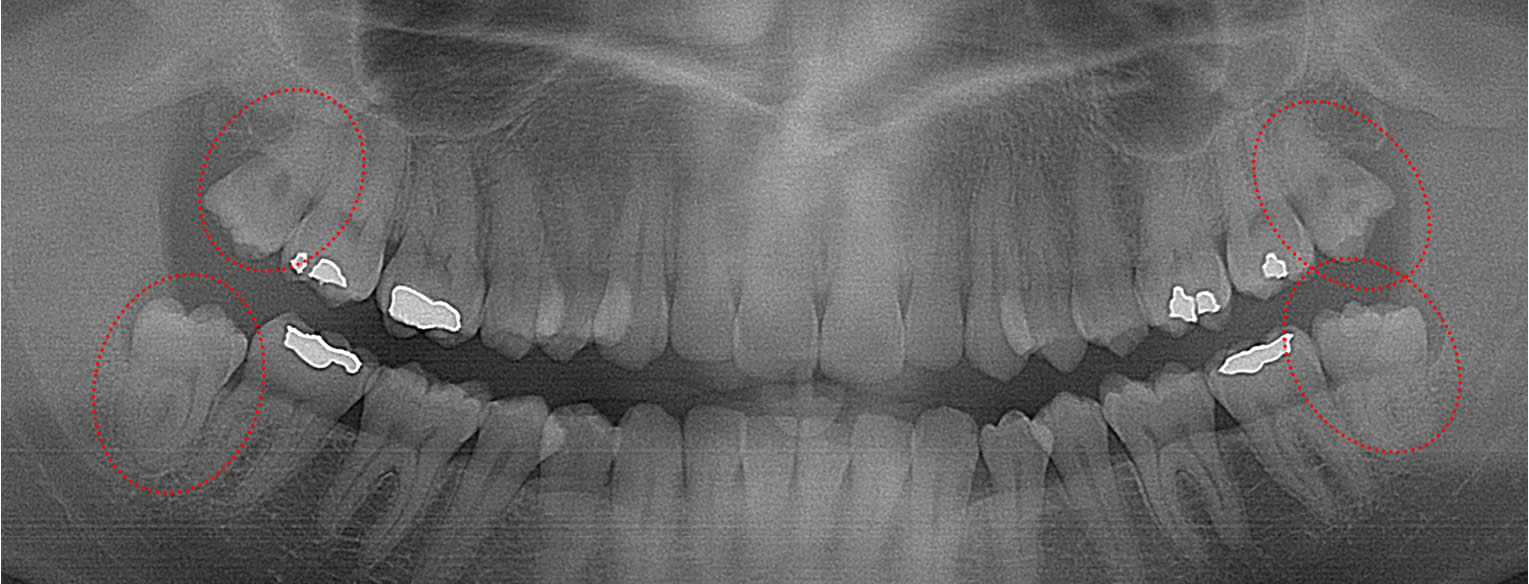
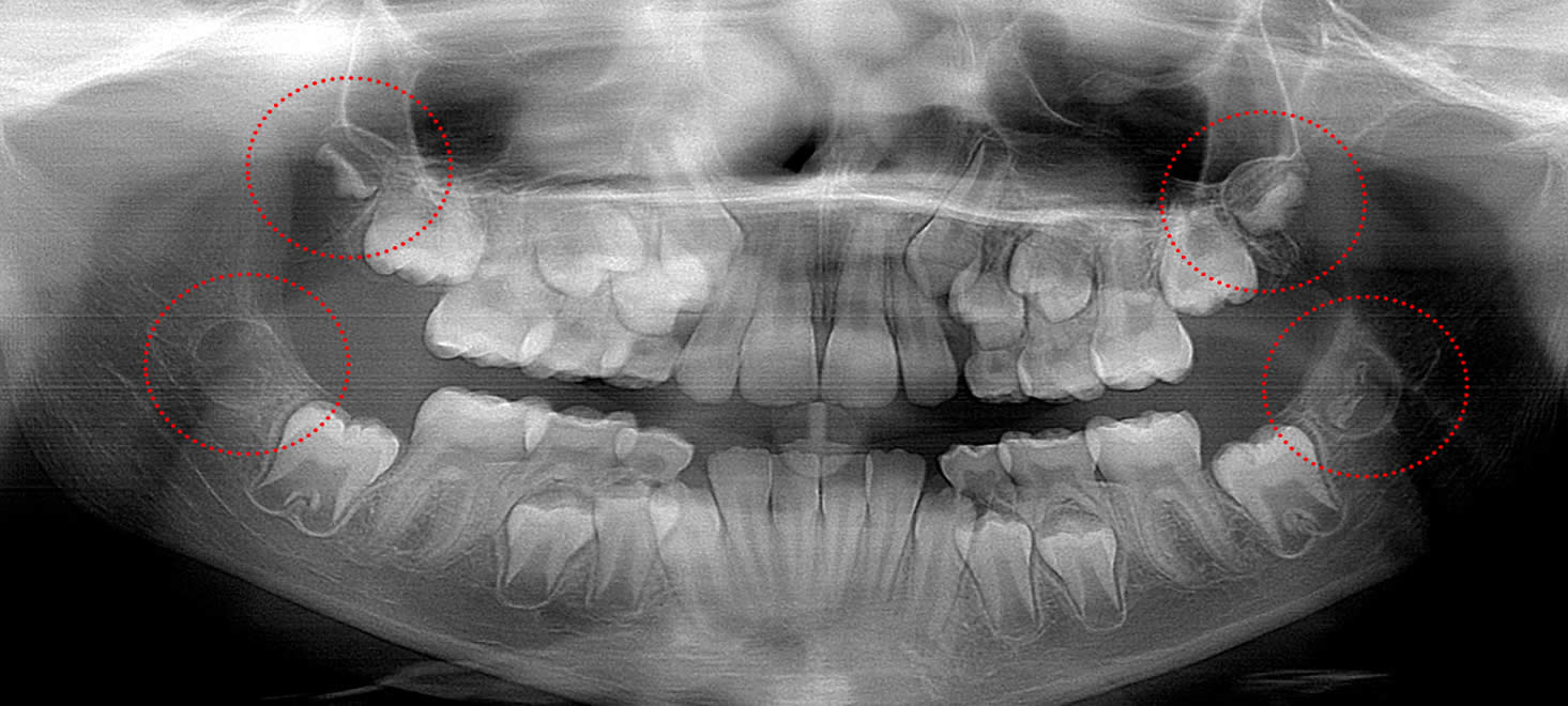
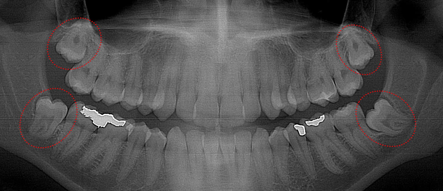
This 19 year old patient has all four 3rd molars present (circled). Only the upper left 3rd molar has fully erupted. The lower left 3rd molar is partially exposed and decaying while the lower right soft tissue impacted, both requiring extraction. Note the double crown on the upper right third molar.
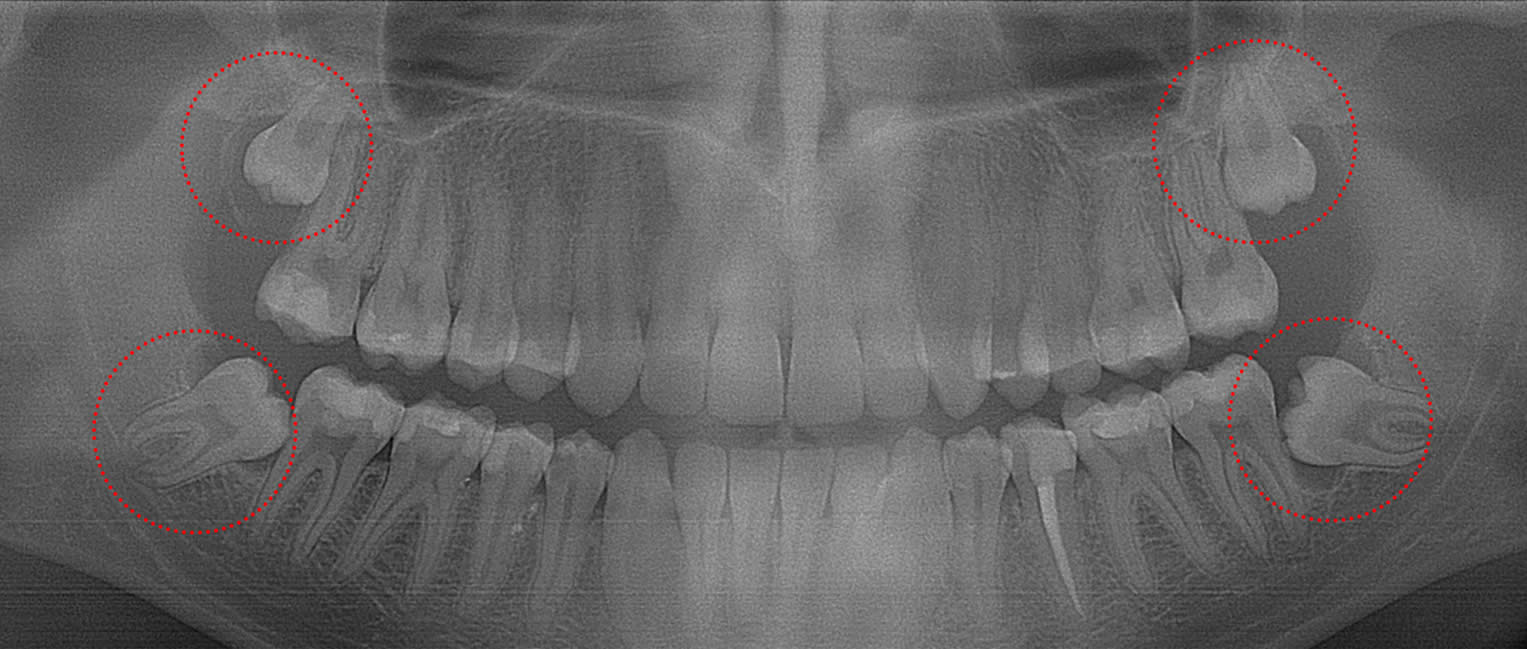
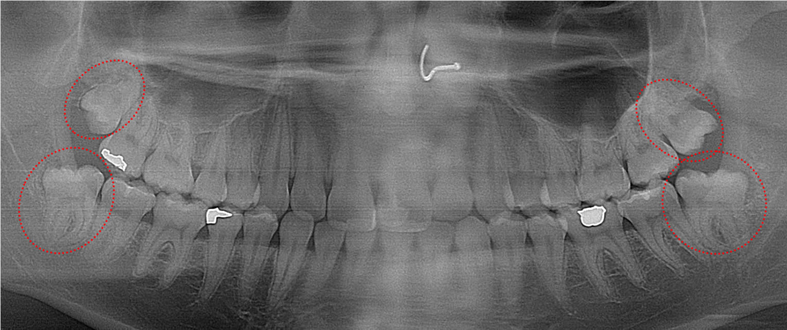
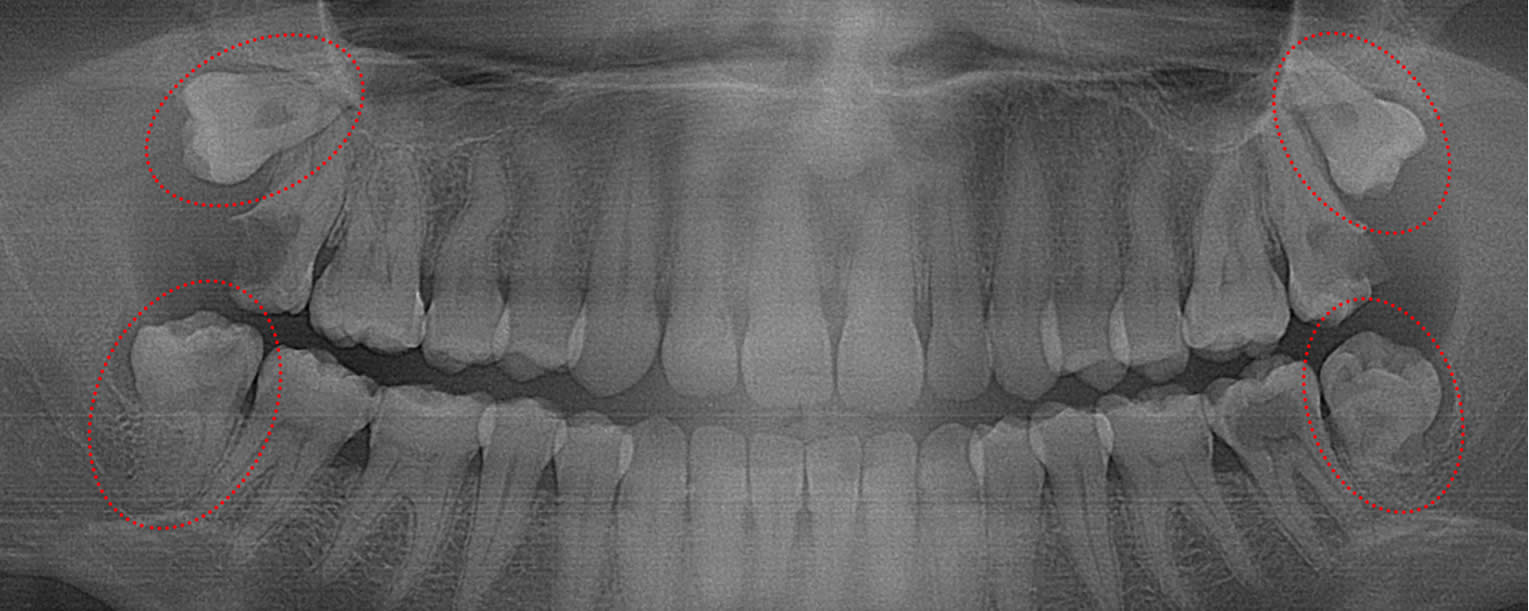
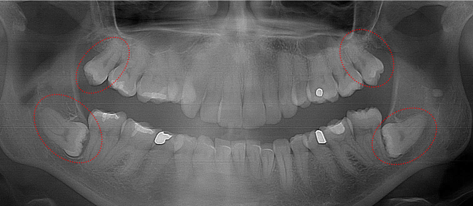
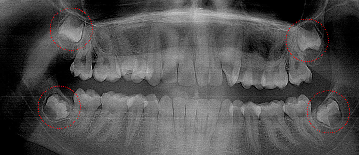
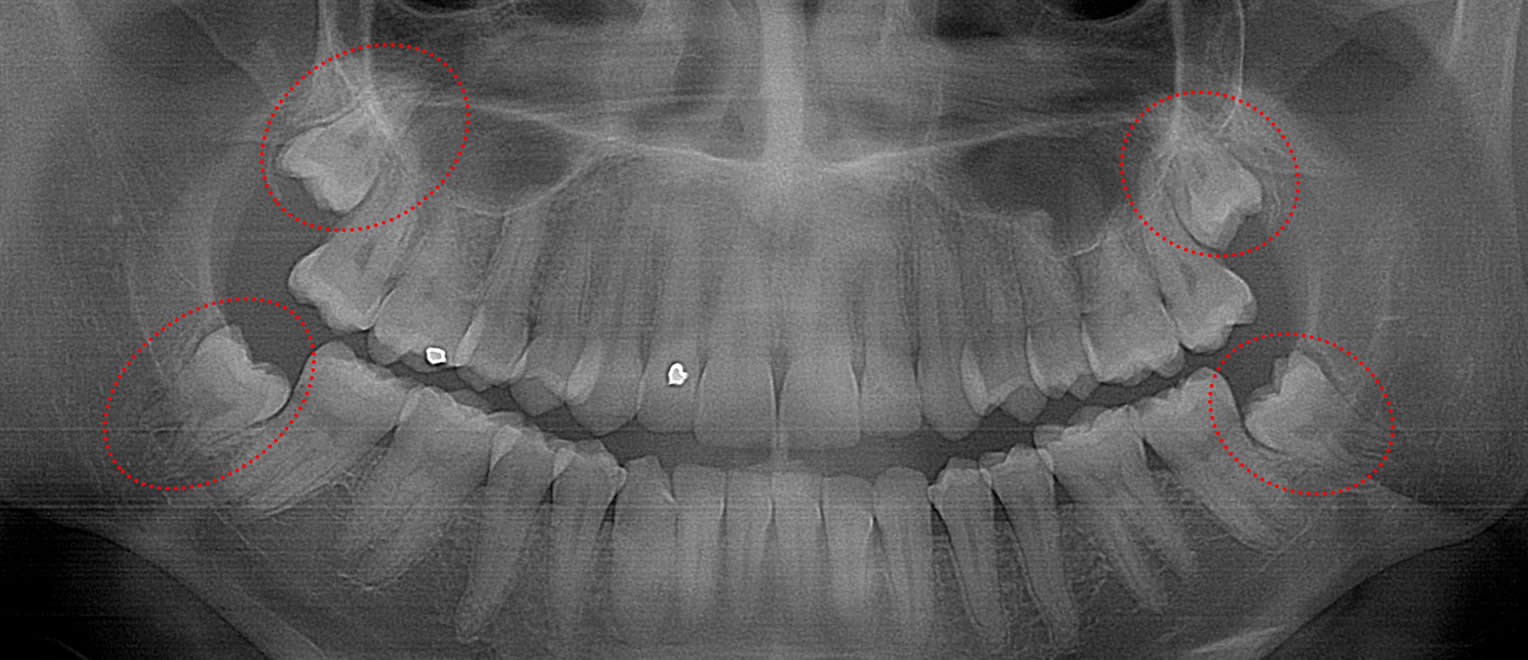
This 19 year old patient has all four 3rd molars present (circled). The roots are +90% formed. Both lower 3rd molars are impacted against the 2nd molars with no chance of further eruption and a +60% probability of decaying before age 30. The patient presented with pain and infection around both lower 3rd molars, requiring immediate extraction
This 34 year old patient has all four 3rd molars present (circled). They are all partially erupted and decaying. The prognosis for all four 3rd molars is very poor and extraction is recommended.
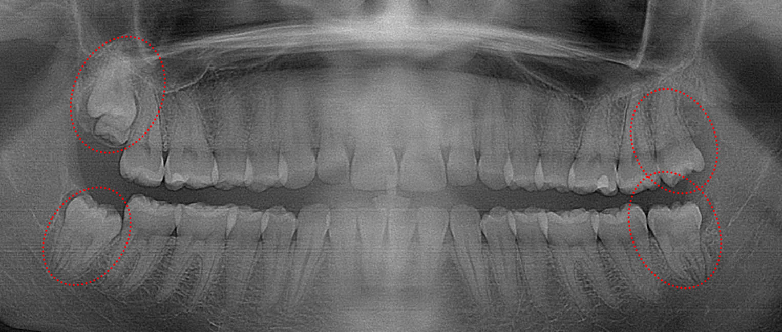
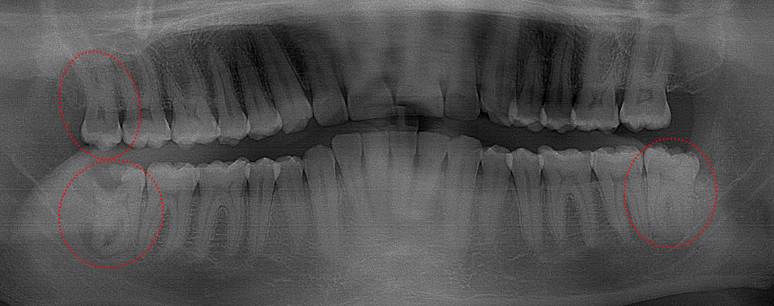
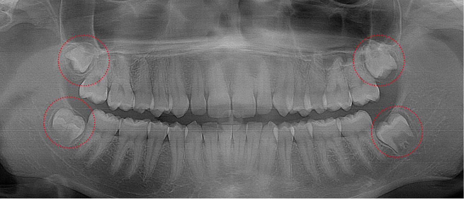
This 21 year old patient has all four 3rd molars present (circled). The orientation of all four teeth is very poor with the lower teeth tipping posteriorly into the main bone. These are very difficult to extract because bone must be removed and each tooth sectioned to get them out.
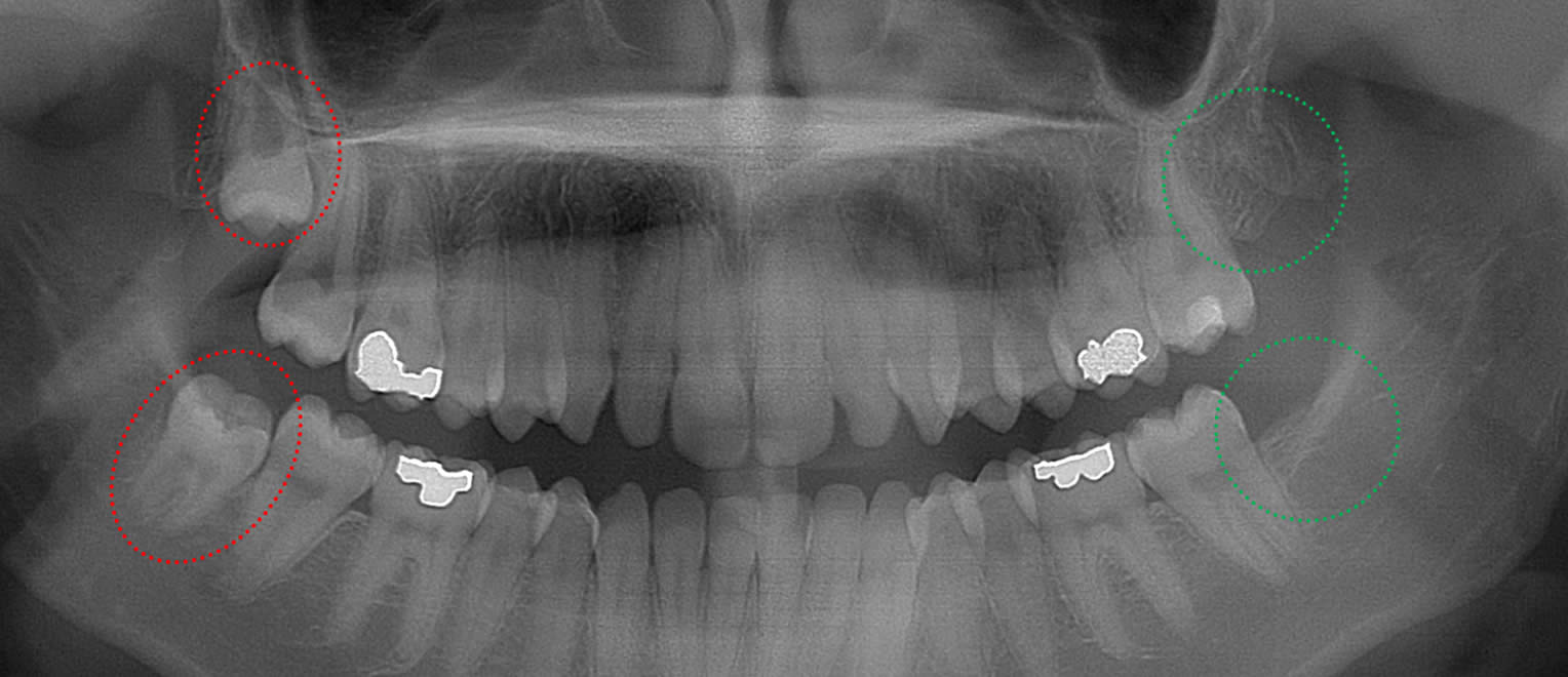
This 33 year old patient has three 3rd molars present (circled). The lower right 3rd molar is badly decayed with a severe abscess at the root tips. The lower left 3rd molar is starting to decay. Extraction of all three 3rd molars is recommended because these are the most commonly decayed teeth.
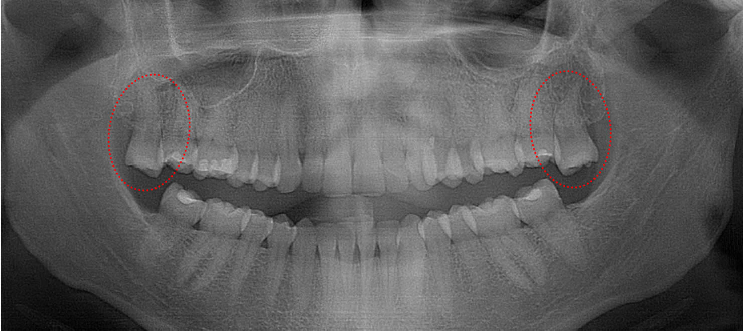
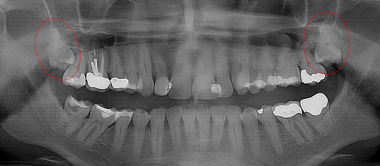
This 29 year old patient had her two impacted lower 3rd molars extracted 3 years ago…but not her uppers. Both upper third molars (circled) have hyper-erupted and the patient is physically biting down on her gum tissue on both sides and suffers from painful open sores.
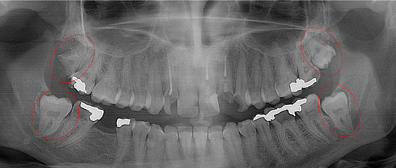
This 29 year old patient has all four 3rd molars present (circled). The upper left 3rd molar is badly decayed and the prognosis is poor for the remaining three because they are partially exposed and will not erupt any further. Extraction of all four 3rd molars is recommended.
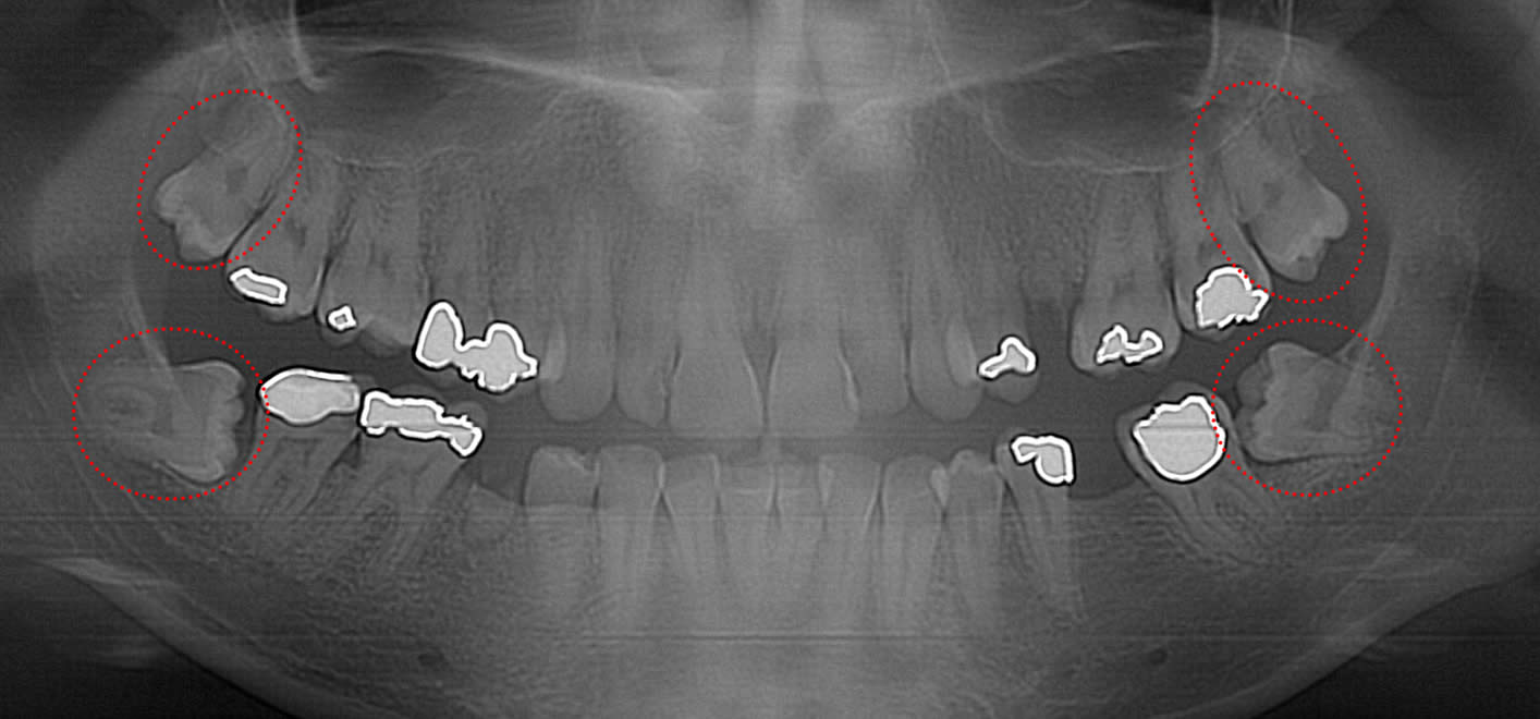
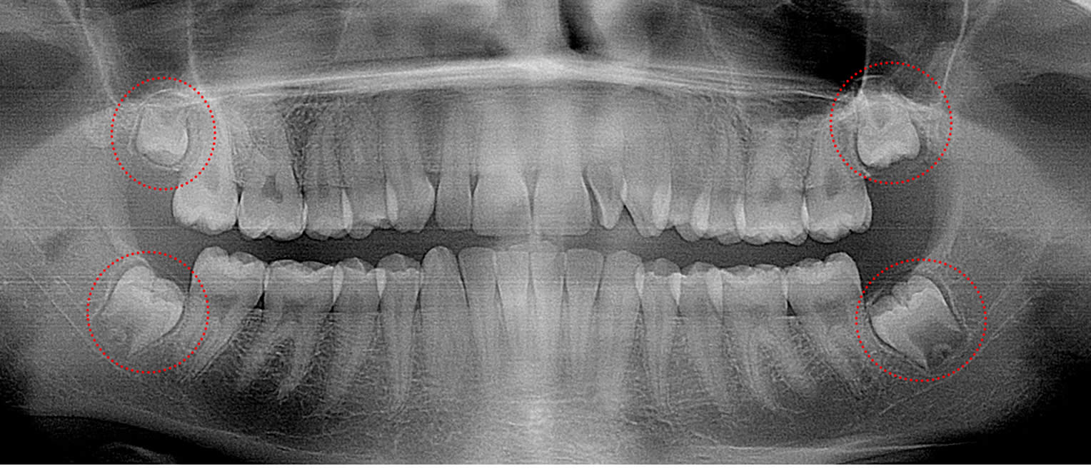
This 26 year old patient has all four 3rd molars present (circled). Given the extensive decay present on adjacent teeth and the highly tipped orientation of the 3rd molars, all four 3rd molars are recommended for immediate extraction before decay can be treated.
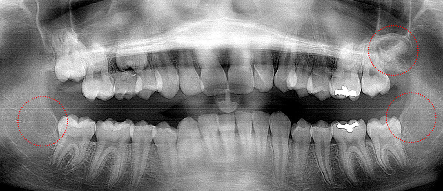
This 26 year old patient has all four 3rd molars present (circled). The upper right 3rd molar is badly decayed and the other three are partially erupted with signs of decay forming. Immediate extraction of all four is recommended.
This 48 year old patient has both upper 3rd molars present (circled). There is a severe gum infection around both 2nd and 3rd molars as a result of the 3rd molars pushing forward into them. Extraction of 2nd and 3rd molars is recommended in order to save the 1st molars.
This 36 year old patient has all four 3rd molars present (circled). The uppers are hyper-erupting and decaying. The lower 3rd molars are horizontally impacted and show no signs of infection…but it is very likely that the adjacent 2nd molars will become badly infected in the next 10 years.
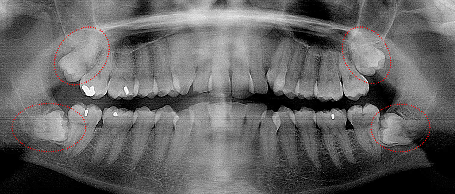
This 32 year old patient has all four 3rd molars present (circled). She presents with pain involving the lower 3rd molar with swelling over it. Immediate extraction of all four 3rd molars is recommended.
This 29 year old patient has all four 3rd molars present (circled) and presented with pain and infection involving both lower 3rd molars. Extraction is required, but there will be huge boney defects distal to the 2nd molars, making the prognosis very poor for the 2nd molars.
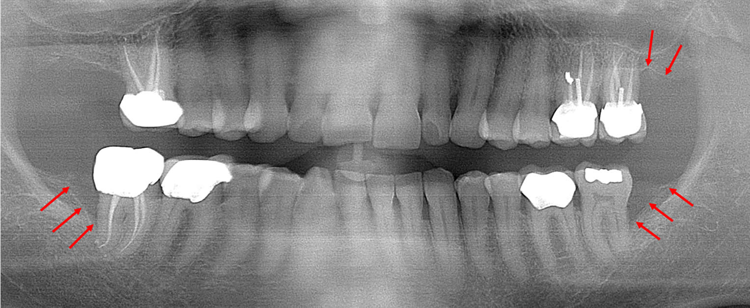
This 48 year old patient had all four 3rd molars extracted in his late 20’s. All four sites healed poorly and chronically infected bony defects remained. The upper right 2nd molar was extracted two years ago as a result of this chronic infection. The upper left 2nd molar must now be extracted because of a chronic periodontal infection moving around that tooth. Both lower molars are chronically infected and have a poor prognosis.
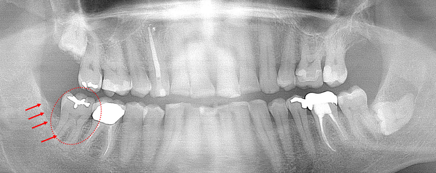
This 52 year old patient had the lower right 3rd molar extracted 6 years earlier due to a chronic infection. The boney defect (arrows) posterior to the 2nd molar (circled) is a site with a chronic gum infection present. Repeated cleaning is necessary to prevent further bone loss around the 2nd molar. Note the other two 3rd molars laying in the bone.
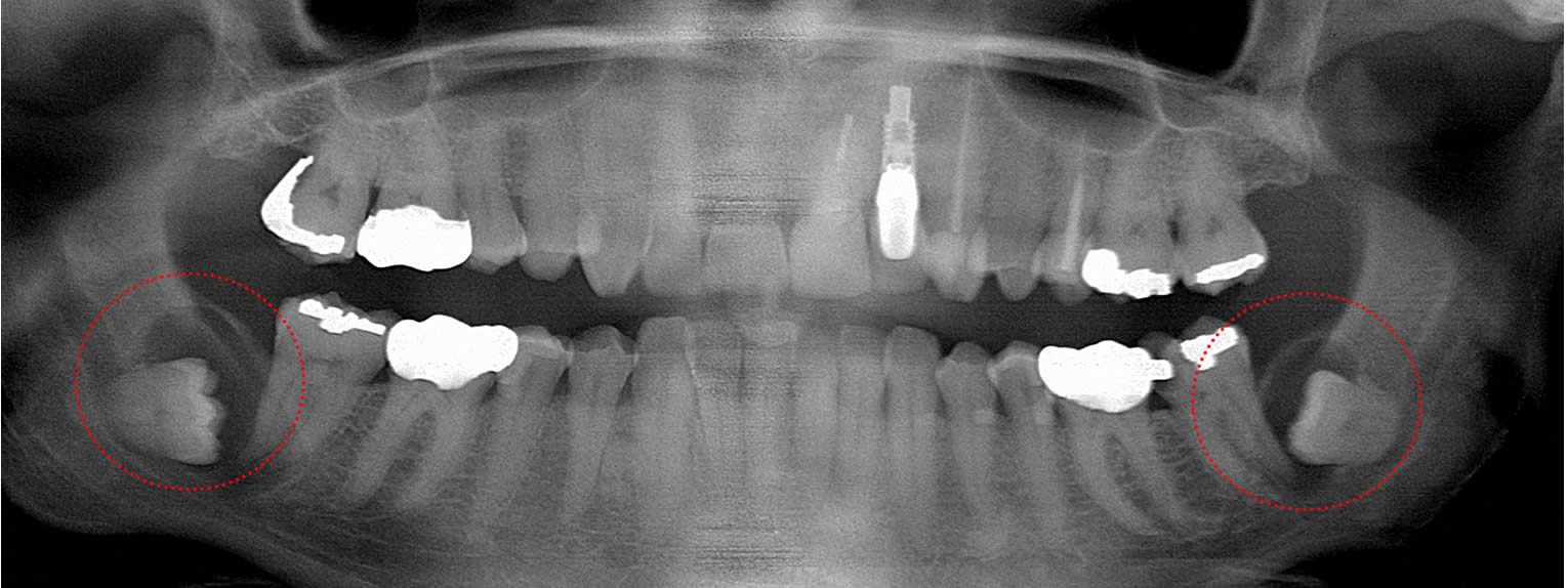
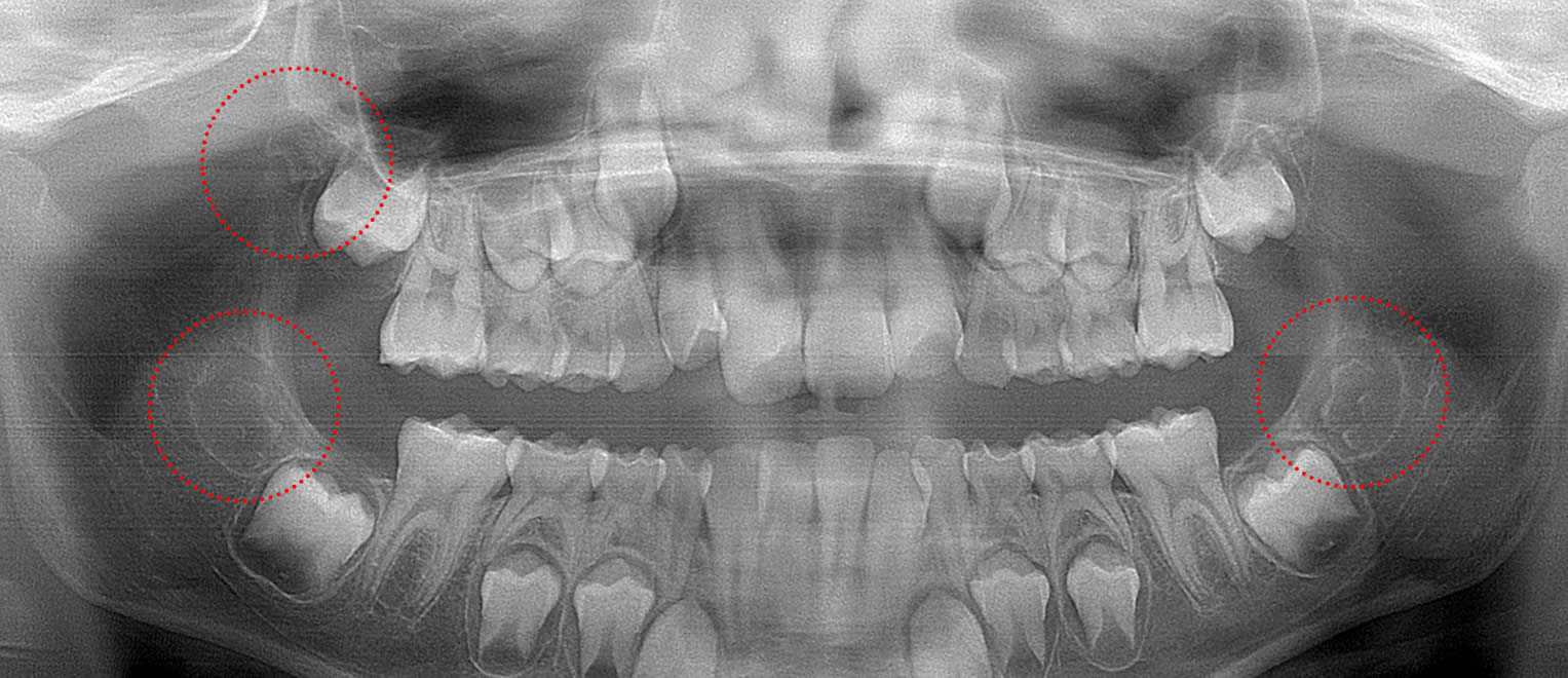
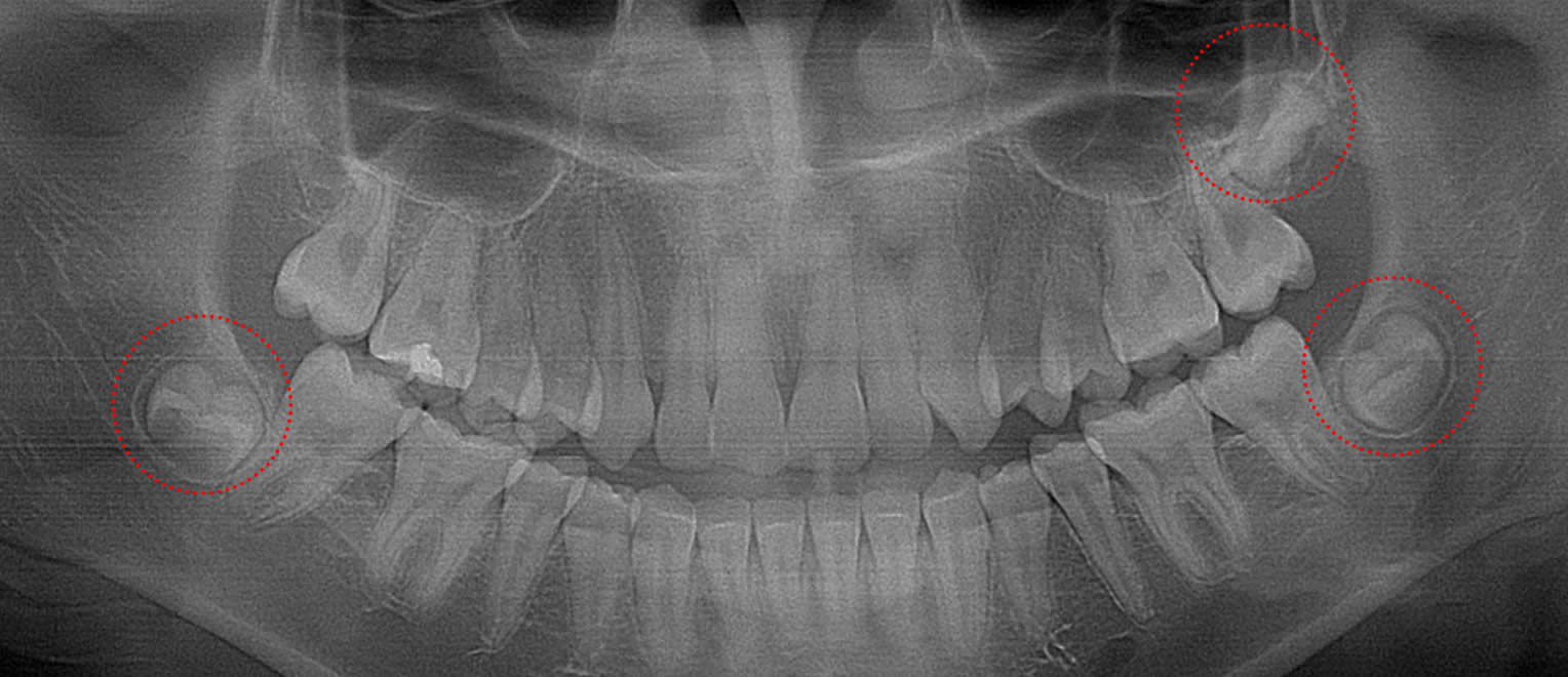
This 55 year old patient has bilateral mandibular 3rd molars present with cysts (circled) forming around them. Failure to have the 3rd molars and the expanding cysts removed will eventually lead to fractures in the mandible as the bone thins.
Zero3™ TBA (3rd molar Tooth Bud Ablation) takes advantage of the fact that tooth buds that form wisdom teeth start growing much later than any other tooth bud. Wisdom tooth formation is first detectable in children around age 6. The following x-rays show the progress of the first-detectable wisdom tooth bud as it grows, starts forming initial tooth structure around age 9-12 and then grows into a nearly fully formed tooth after age 14 before erupting. This all occurs before any significant pathology occurs once the tooth becomes fully formed and then tries to erupt through gum tissue into the oral cavity.
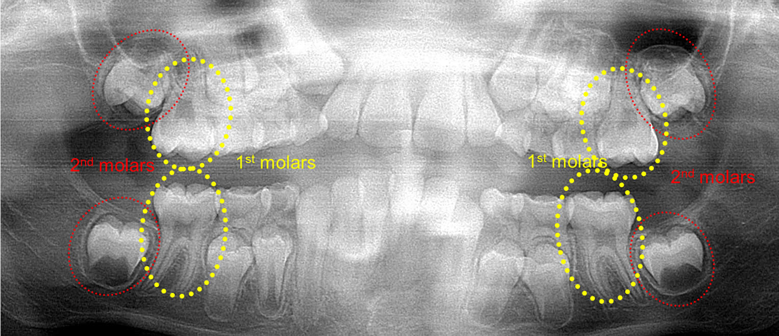
This 8 year old patient has 4 permanent 1st “6 year” molars fully erupted (circled). All 4 permanent 2nd molars are deep in the jaw on both arches and are just starting root formation (circled). There are no 3rd molar tooth buds evident below, but natural 3rd molar agenesis that occurs 7% of the time cannot be confirmed until age 14.
This 9-year old patient has three 3rd molars forming (circled). The cusp tips are barely detectable, indicating that enamel formation has just recently started. It is not certain that the upper left tooth bud will not form.
This 10 year old patient has four 3rd molar tooth buds present (circled) with three of them in the very early stages of enamel formation. The lower right 3rd molar tooth bud may have just a trace of enamel formed.
This 13 year old patient has two lower arch 3rd molar tooth buds present with no enamel formation (circled). The upper left 3rd molar tooth bud (circled) has formed extensive enamel. There is no evidence of an upper right wisdom tooth bud.
This 13 year old patient has all four 3rd molars present (circled). The lower 3rd molars are tipped laterally and slightly towards the midline enough to make normal eruption uncertain, even if there will be adequate space present to erupt into in 2-4 years.
This 14 year old patient has three 3rd molars showing mid-stage enamel formation (circled). Root formation is still 2-4 years away. There is no way to assess whether this patient will have adequate room for them to erupt normally.
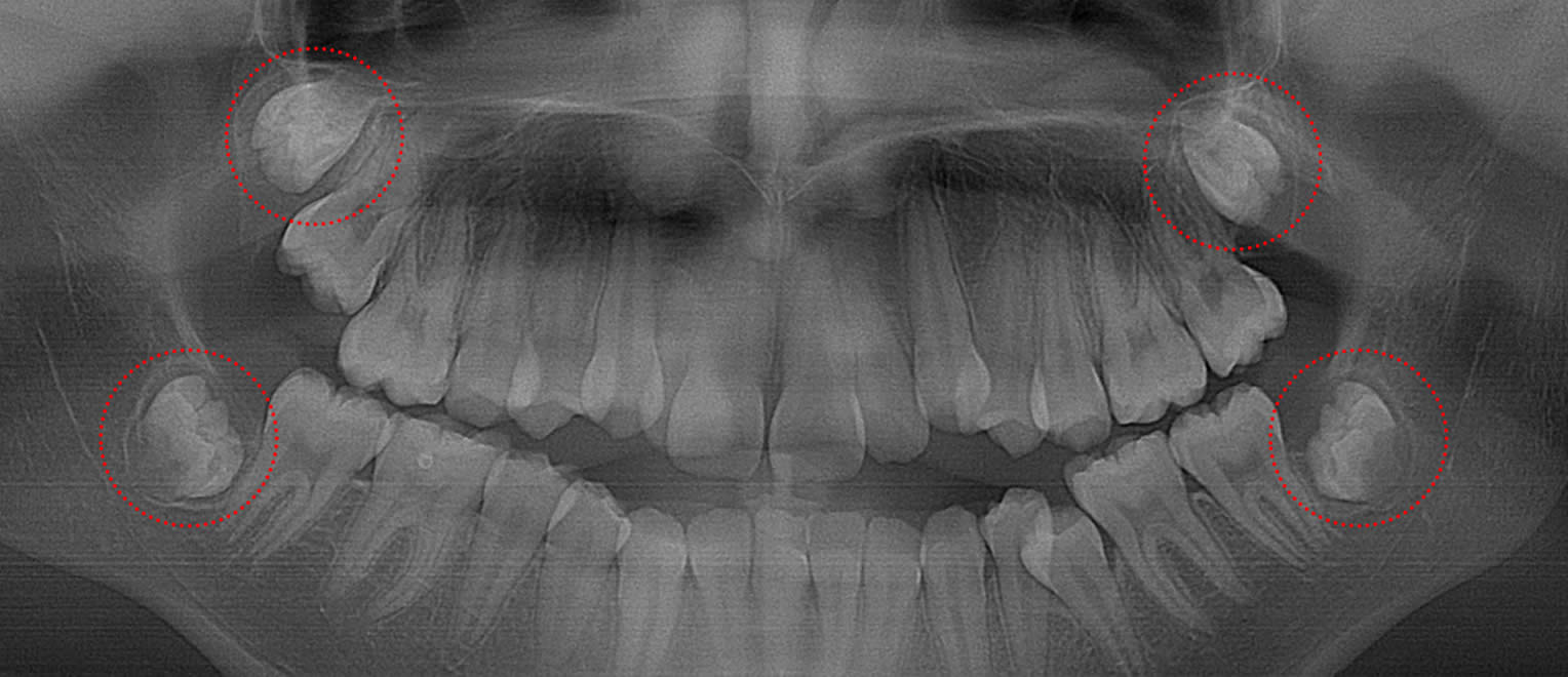
This 15 year old patient has four 3rd molars developing (circled). Enamel formation is nearly complete and the first signs of root formation are evident. There may be adequate room for normal eruption, but it is too early to tell with certainty.
This 17 year old patient has all four 3rd molars present (circled). Root formation is just starting with eruption still 2-4 years away. There does not appear to be space for normal eruption and both right molars may be tipped laterally, resulting in a boney impaction or ectopic eruption.
This 15 year old patient has all four 3rd molars present (circled). Roots are approximately 1/3rd formed and they are oriented well with a good possibility of normal eruption 2-4 years from now. However, there is a high chance they will be soft-tissue impacted and decay as a result.
This 18 year old patient has only two 3rd molars present (circled). Compare the solid bone behind the upper left 2nd molars (circled) where no wisdom tooth formed to the missing bone on the distal of the upper right 2nd molar.
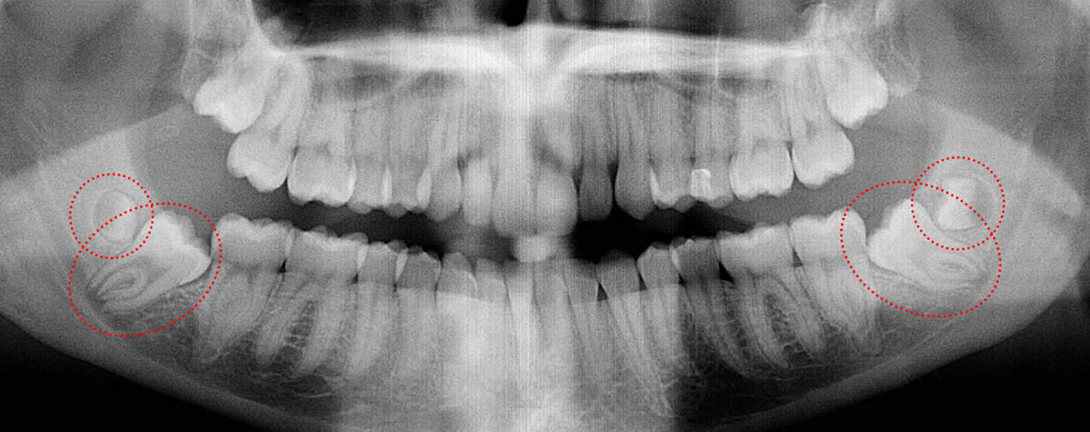
This 21 year old patient has all four 3rd molars present (circled). The roots are approximately 2/3rds formed. The lower right 3rd molar cannot erupt any further; it is distally tipped into the ramus of the mandible. The lower left 3rd molar likely will not erupt any further; it is pushing into the undercut of the distal of the 2nd molar.
This 19 year old patient has all four 3rd molars present with root formation approximately 3/4ths completed (circled). There appears to be good orientation and space, but they may become soft tissue impacted and decay as a result. There is no way to predict the outcome.
How would you like to be this 21 year-old patient? Count the number of “3rd” molars (circled) on the lower arch…ouch…there are supranumery wisdom teeth present!